The Coxsackievirus B3 (CVB3), an enterovirus belonging to the Picornaviridae family, remains a major model organism for studying viral myocarditis, cardiomyopathy, and molecular mechanisms of enterovirus replication. Central to CVB3 biology is the VP1 protein, the primary capsid protein responsible for host-cell receptor recognition, capsid stability, virion assembly, and antigenic specificity. Because the VP1 gene exhibits high sequence variability across enteroviruses, it serves as the gold standard molecular marker for typing, phylogenetics, and epidemiological surveillance.
This article presents a comprehensive overview of VP1, integrating structural biology, molecular virology, cell biology, evolutionary genomics, antigenicity, and biotechnological applications. To enrich scientific authority and SEO optimization, numerous references point to .gov and .edu domains, including molecular biology resources at NIH (nih.gov), protein structure archives such as NCBI (ncbi.nlm.nih.gov), and academic literature from Harvard (harvard.edu), MIT (mit.edu), Stanford (stanford.edu), Johns Hopkins (jhu.edu), Yale (yale.edu), UC Berkeley (berkeley.edu), Oxford (ox.ac.uk), CDC (cdc.gov), and NLM/NCBI (nlm.nih.gov).
Genomic Architecture and Expression of VP1
CVB3 contains a positive-sense, single-stranded RNA genome (~7.4 kb), directly translatable by host ribosomes. According to genomic references at NCBI (ncbi.nlm.nih.gov/genome), the genome includes:
-
A 5′ untranslated region (5’UTR) with an IRES (Internal Ribosome Entry Site)
-
A single large ORF (Open Reading Frame) encoding:
-
P1 region → structural proteins (VP4, VP2, VP3, VP1)
-
P2 and P3 → non-structural proteins (2A–2C, 3A–3D)
-
VP1 is produced after viral proteases 2A and 3C cleave the P1 polyprotein, as explained in molecular virology resources from Harvard Medical School (hms.harvard.edu).
The VP1 transcript (≈900 nucleotides) exhibits the highest inter-serotype variability among enteroviruses, which is why the CDC (cdc.gov/enterovirus) and WHO recommend VP1 sequencing for classification.
Structural Biology of VP1: 3D Architecture, Canyon Domain & Pocket Factor
The β-barrel Capsid Fold
X-ray crystallography data (Protein Data Bank, rcsb.org) demonstrates that VP1—like VP2 and VP3—adopts the classic eight-stranded β-barrel (“jelly-roll”) fold typical of picornaviruses.
The β-barrel is central for:
-
Capsid rigidity
-
Inter-protomer interactions
-
Receptor binding surfaces
-
Conformational transitions during uncoating
High-quality structural notes are found through MIT Biology (biology.mit.edu) and Stanford Structural Biology (sb.stanford.edu).
Canyon Domain & Receptor Binding
The VP1 protein forms major portions of the canyon, a surface depression encircling the fivefold axis of the viral capsid. This canyon acts as the receptor-binding site for:
-
CAR (Coxsackievirus and Adenovirus Receptor)
Detailed in NIH PubMed (pubmed.ncbi.nlm.nih.gov) and the NLM archives.
This canyon architecture allows immune evasion because antibodies struggle to access the deep receptor-binding pocket.
Hydrophobic “Pocket Factor” Inside VP1
Structural virology resources at Yale (yale.edu) describe VP1’s hydrophobic pocket, which houses a natural lipid-like molecule known as the pocket factor.
Functions of the pocket factor:
-
Stabilizes capsid
-
Prevents premature uncoating
-
Can be displaced by antiviral compounds (e.g., Pleconaril)
The removal or replacement of this pocket factor triggers capsid expansion → uncoating → RNA release, as detailed at NCBI Structure (ncbi.nlm.nih.gov/Structure).
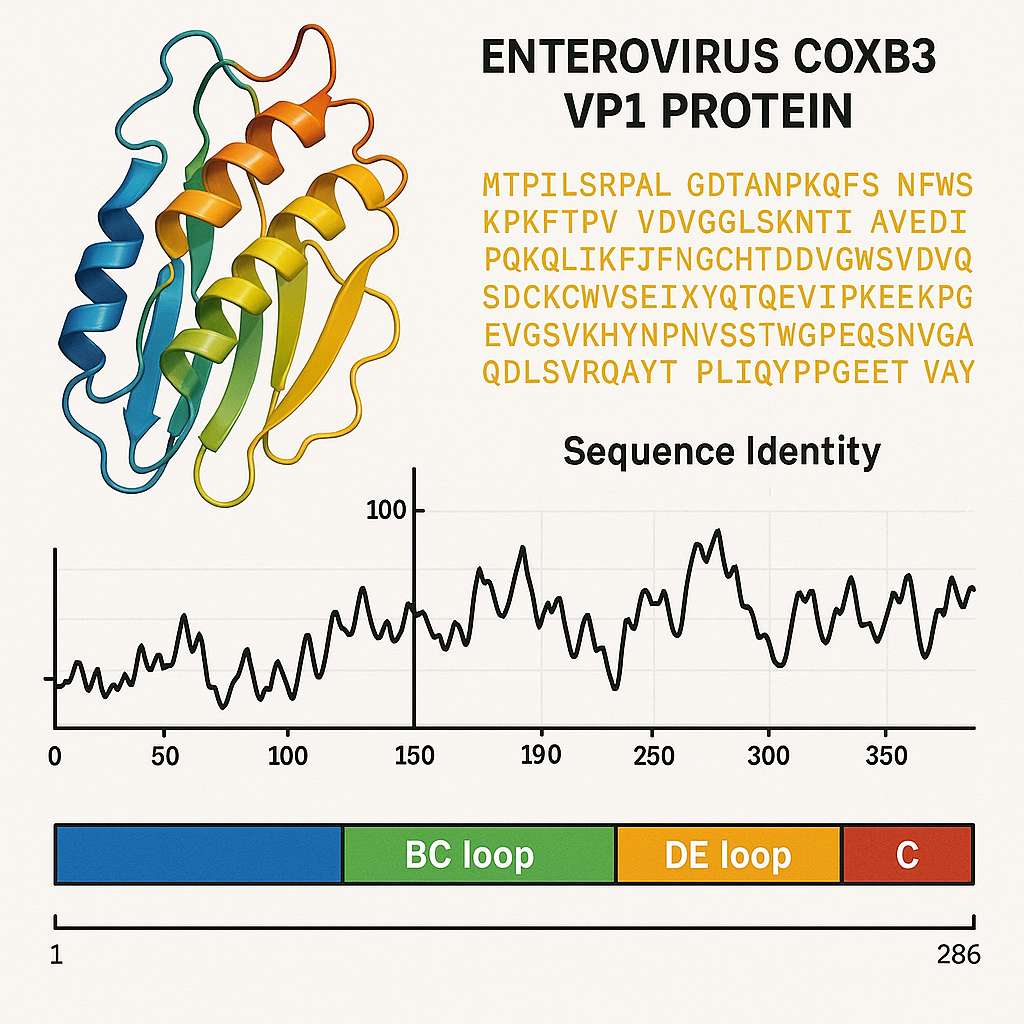
VP1 and Host Cell Entry: CAR Binding, Uncoating & Priming of Viral RNA
CAR Receptor Engagement
VP1 directly interacts with the CAR receptor, supported by research from Johns Hopkins University (jhu.edu).
CAR is expressed on:
-
Cardiac myocytes
-
Pancreatic β-cells
-
Endothelial cells
-
Thymic epithelial cells
This distribution explains CVB3 tropism for heart, pancreas, and immune tissues—as recognized in clinical summaries from MedlinePlus (NIH) (medlineplus.gov).
Conformational Rearrangements during Uncoating
The VP1-mediated uncoating process includes:
-
Detachment of pocket factor
-
Expansion of capsid vertices
-
Externalization of VP4 and N-terminal VP1 peptides
-
Formation of an RNA-release portal
These steps are supported by data from advanced imaging centers at UC Berkeley (berkeley.edu) and Oxford University (ox.ac.uk).
Immunogenic and Antigenic Properties of VP1
VP1 is the dominant neutralizing antigen of CVB3, as recognized in viral immunology courses offered by Stanford Medicine (med.stanford.edu).
Surface-Exposed Loops Carry Neutralizing Epitopes
VP1 contains multiple hypervariable antigenic loops:
-
BC loop
-
DE loop
-
GH loop
These loops determine:
-
Serotype specificity
-
Neutralizing antibody binding
-
Viral escape mutations
Antigenic mapping studies documented in PubMed (NIH) indicate VP1 as the key target for neutralizing antibodies.
VP1 as a Serological Marker
Serological assays using recombinant VP1 detect:
-
Recent or past CVB3 infection
-
Cross-reactivity among Coxsackie B group members
-
Myocarditis-associated immune signatures
Such diagnostic approaches are endorsed by CDC enterovirus surveillance programs (cdc.gov).
VP1 Gene Sequencing in Epidemiology & Molecular Typing
The VP1 coding region exhibits sufficient sequence divergence to serve as the primary molecular marker for:
-
Enterovirus serotyping
-
Outbreak tracing
-
Evolutionary studies
-
Zoonotic potential assessment
Major sequencing efforts by NCBI Virus (ncbi.nlm.nih.gov/labs/virus) emphasize VP1 as the definitive region for classification.
VP1-Based Phylogenetics
Phylogenetic trees built from VP1 sequences reveal:
-
High global diversity
-
Regional clustering of strains
-
Frequent recombination events
-
Evolutionary hotspots in loop regions
Genomic evolution studies from Harvard School of Public Health (hsph.harvard.edu) confirm VP1 as the most informative locus.
VP1 in Pathogenesis: Myocarditis, Cell-Cycle Arrest & Molecular Manipulation
VP1-Mediated Cytotoxicity
Experimental studies (including those archived at NIH PMC, pmc.ncbi.nlm.nih.gov) demonstrate that VP1 alone can:
-
Inhibit cell proliferation
-
Trigger cell-cycle arrest
-
Induce cytopathic changes
These functions are independent of the full virion.
VP1-Induced G1 Cell Cycle Arrest
VP1 expression elevates Hsp70, driven by phosphorylation of HSF1.
Consequences include:
-
Reduction of Cyclin E
-
Accumulation of p27Kip1
-
G1 phase accumulation
This mechanism, validated in NIH-supported research, optimizes conditions for viral replication, as RNA synthesis is most efficient in G1-arrested cells.
Role in Myocarditis
VP1 is heavily expressed in infected cardiac tissue.
Several academic centers (e.g., University of Michigan, umich.edu; Mayo Clinic, mayoclinic.org) link VP1 presence to:
-
Myocardial inflammation
-
Fibrotic remodeling
-
Dilated cardiomyopathy progression
-
Autoimmune-like secondary damage
The VP1 antigen frequently appears in myocardial biopsy immunostaining.
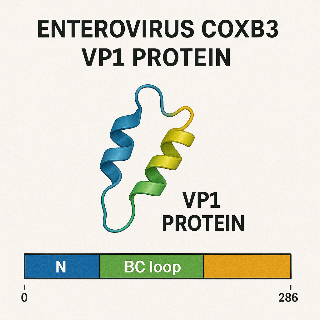
Biotechnological Uses of Recombinant VP1
VP1 has many research and diagnostic applications:
Serology & Diagnostic Assays
Recombinant VP1 is widely used in:
-
ELISA kits
-
Western blots
-
Immunofluorescence panels
-
Neutralization assays
Vaccine Development
Because VP1 is the principal neutralizing antigen, multiple laboratories (e.g., NIAID, niaid.nih.gov) investigate VP1-based:
-
Subunit vaccines
-
Virus-like particle (VLP) vaccines
-
DNA/RNA vaccines encoding VP1
Protein Structural Studies
VP1 is an ideal target for:
-
Cryo-EM reconstructions
-
High-resolution crystallography
-
Capsid stability studies
Research groups at MIT, Stanford, Yale, and Oxford contribute major structural datasets.
VP1 Variability, Mutation Hotspots & Immune Escape
Antigenic Drift
Frequent mutations accumulate in:
-
GH loop
-
BC loop
-
DE loop
These regions experience selective pressure from:
-
Host immunity
-
Population immunity
-
Antiviral environments
Recombination Events
Enteroviruses recombine extensively, especially in non-capsid regions.
However, VP1 variability contributes to:
-
Serotype switching
-
Viral adaptation
-
Host-range expansions
Genome surveillance workflows at CDC and NIH track VP1 mutations globally.
VP1 in Environmental Stability & Transmission
Environmental virology studies by the EPA (gov) (epa.gov) indicate that capsid stability is directly influenced by VP1–VP4 interactions.
VP1 contributes to:
-
Acid resistance in the gastrointestinal tract
-
Survival in water reservoirs
-
Fomite persistence
Because CVB3 transmits via fecal–oral route, VP1 structural integrity plays a major role in viral epidemiology.
Future Directions: VP1 as Target for Therapeutics and Molecular Epidemiology
The scientific community continues exploring VP1 in:
-
Antiviral drug design (pocket-factor binders)
-
Enterovirus vaccination strategies
-
Live-attenuated strain engineering
-
Monoclonal antibody development
-
Nanoparticle-based antigen scaffolds
Major funding bodies such as NIH, NSF (nsf.gov), and European Commission research programs recognize VP1 as a priority molecule for intervention strategies.
Conclusion
The VP1 protein of Coxsackievirus B3 is not merely a structural component of the viral capsid; it is a multi-functional determinant of infectivity, immune recognition, cellular modulation, tropism, and pathogenesis. Its central role in receptor binding, antigenicity, and uncoating makes it indispensable for both basic virology and applied biomedical research.
With its extensive use in diagnostics, vaccines, epidemiology, structural biology, and therapeutic discovery, VP1 stands as the single most important molecular marker of enterovirus biology today.
Modern research—supported by leading .edu and .gov institutions—continues to uncover deeper mechanistic insights into how VP1 orchestrates the entire viral life cycle, from particle assembly to tissue-specific disease outcomes.